Skip to Content

Health Encyclopedia

X-ray of rheumatoid arthritis in the feet

« Previous Page En Español

X-ray of rheumatoid arthritis in the feet

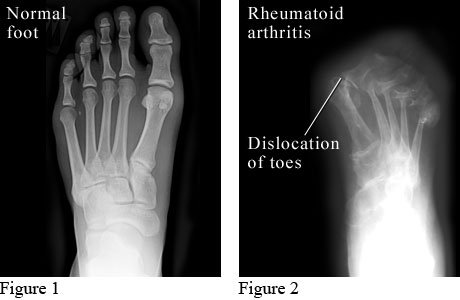
X-ray images of a normal foot and a foot with rheumatoid arthritis

Figure 1 courtesy of Intermountain Medical Imaging, Boise, Idaho. Figure 2 courtesy of Paul Traughber, MD, Boise, Idaho.

The X-ray on the left shows a normal foot.

The X-ray on the right shows a foot with advanced rheumatoid arthritis. Cartilage and bone are worn away, and the bones of the toes have moved out of their normal positions.

Current as of: July 10, 2023

Author: Healthwise Staff
Clinical Review Board
All Healthwise education is reviewed by a team that includes physicians, nurses, advanced practitioners, registered dieticians, and other healthcare professionals.

This information does not replace the advice of a doctor. Healthwise, Incorporated, disclaims any warranty or liability for your use of this information. Your use of this information means that you agree to the Terms of Use. Learn how we develop our content.

© 1995-2024 Healthwise, Incorporated. Healthwise, Healthwise for every health decision, and the Healthwise logo are trademarks of Healthwise, Incorporated.

This information does not replace the advice of a doctor. Healthwise, Incorporated, disclaims any warranty or liability for your use of this information. Your use of this information means that you agree to the Terms of Use. Learn how we develop our content.